CT is much faster than MRI making it the study of choice in cases of trauma and other acute neurological emergencies. A brain aneurysm is a weak area in the wall of a blood vessel in the brain.

Mri Vs Ct Cat Scan Which Is Best For My Brain Imaging Needs
Learn more Cognitive Care Planning.

. Examine the skull or blood vessels with a CT scan. The scan can show if theres a fracture or bleeding. The forebrain the midbrain and the.
Diagnostic angiogram During this procedure a catheter is used to look for abnormalities in the blood vessels using a rapid set of x-rays and time-lapse photography. Amen Clinics is different because we use a brain imaging diagnostic tool called SPECT Single Photon Emission Computed Tomography to help accurately identify underlying brain issues. There are many different methods to go about capturing information on brain structures and functions.
Pain scale results can help guide the diagnostic process track the progression of a condition and determine how effective a treatment is. If such a resection. Your doctor will start by requesting your medical history and reviewing your symptoms.
The brain can be divided into three basic units. At this time we do not have firm evidence as to the precise areas of the brain that cause ADHD behaviors. A CT scan takes pictures to create images of the brain.
No it isnt. Functional magnetic resonance imaging fMRI can detect changes in blood flow and oxygen levels that result from your brains activity. If you arent sure.
Cerebral arteriogram also called a cerebral angiogram. CT can be obtained at considerably less cost than MRI. Medications to limit secondary damage to the brain immediately after an injury may include.
Although it resembles a computer mouse the device is backed by. Stethoscopes are probably the most recognizable of all medical diagnostic tools. Brain Gauge is a cognitive assessment tool that uses touch-based sensory testing to measure brain health.
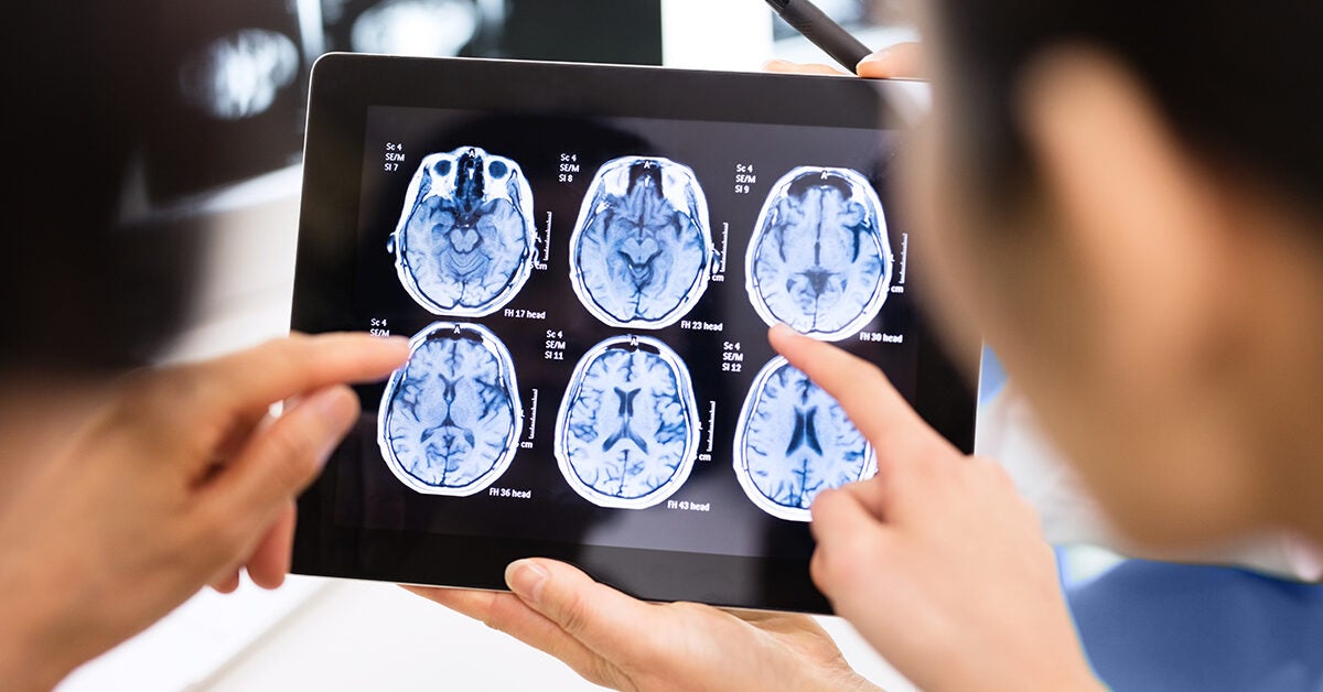
The decision to order contrast-enhanced CT is based on the clinical question being asked. The three most common and most frequently used measures are. An MRI creates clear images of brain tissue.
If you have a brain tumor a head injury or an aneurysm a CT scan is the best way to examine your injury. Common tools used for diagnosing brain cancer include. Contrast agents are used to differentiate between organs and improve lesion.
Currently such increases are detected directly using a sensor that. People whove had a moderate to severe. A range of tools to help physicians.
Tool is used to communicate the level of consciousness LOC of patients with an acute brain injury The scale was developed to complement and not replace assessments of other. But these scans cannot show if you. They measured the thickness of grey matter in 64 different regions of the brain and used this to develop their own person-based similarity index PBSI for short a number.
The medical history and neurologic examination help doctors determine whether herniation is a risk. Psychometric tests or psychological tests consist of a number of formalized tests that tap nearly every domain of psychological personality. They are used to listen to heart sounds the lungs and even blood flow in the.
They will also perform tests that can help them assess your neurological function. An MRI wont help your doctor diagnose ADHD. X-rays are taken after a special.
Whenever possible a full surgical resection of the cancerous tissue is performed. It can burst and cause a stroke and can even lead to death. Herniation puts pressure on the brain and may be fatal.
Detailed reporting across cognitive domains helps providers choose appropriate next steps and referral pathways. It uses the magnetic field of the scanner to affect. The chapter also provides a conceptual model.
Doctors use imaging testslike CT scans or MRIsto. All the parts of the brain work together but each part has its own special properties. For example doctors use an.
All pain scales help improve. Elevations in intracranial pressure following an accident can lead to brain injury and damage to the spinal cord. A cerebral arteriogram is an x-ray or series of x-rays of the head that shows the arteries in the brain.
This chapter provides an overview of traumatic brain injury TBI including how it is defined its mechanisms of injury and its neuropathology.

Zyto Scan And Not Rose Oil Personal Testimony Interesting Info About The Brain And Sense Of Smell Too Limbic System Brain Anatomy Anatomy

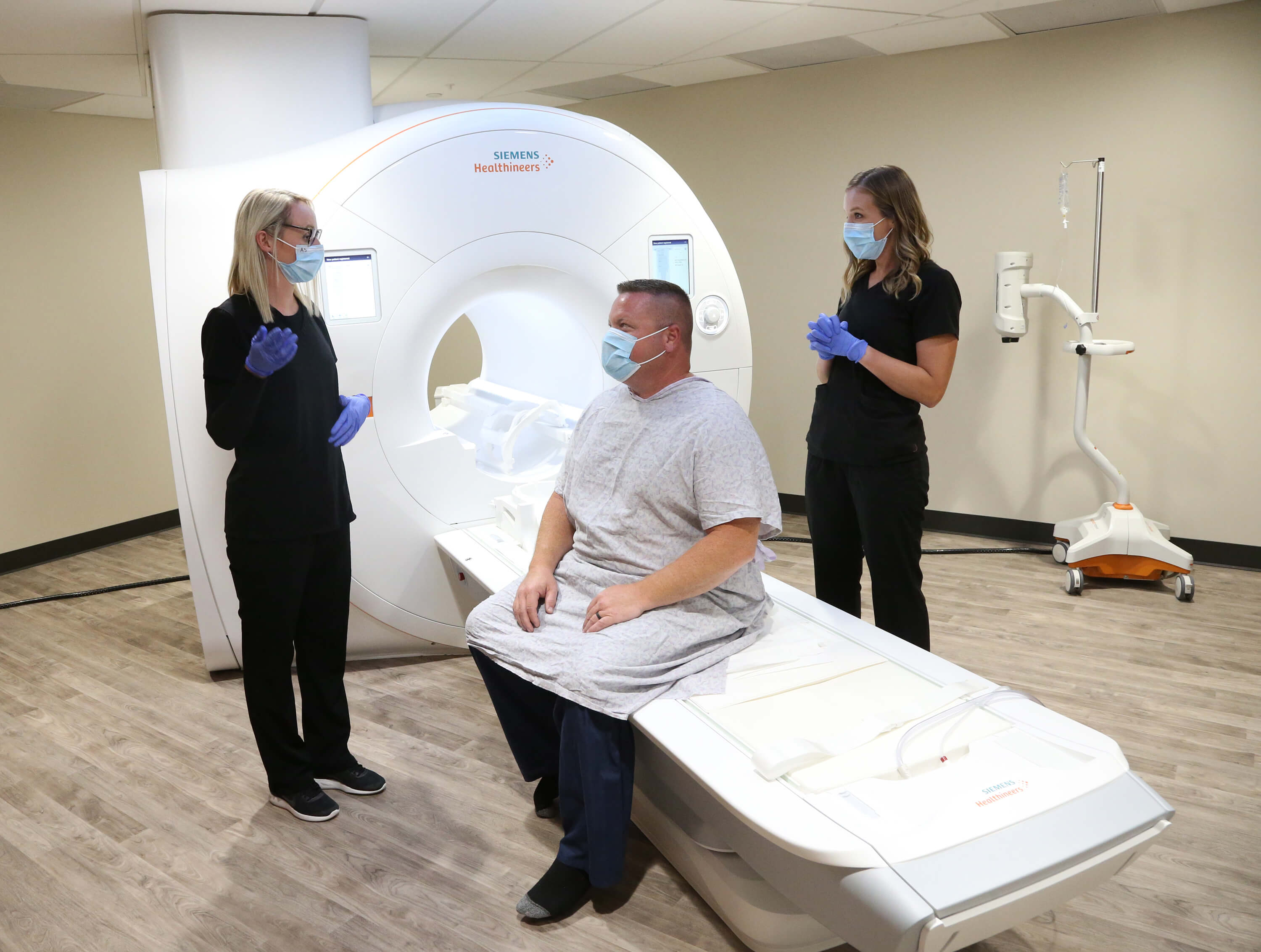
Brain Mri Scan Tesla Mri Machine Echelon Health

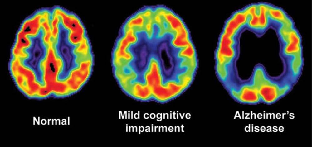
Brain Imaging For Alzheimer S Dementia Pacific Brain Health Center

0 Comments